


by Dr. Beth Johnson
Cause: Mycobacteriosis (commonly referred to as Johne’s disease) is caused by a bacteria Mycobacteria avium subspecies paratuberculosis (often referred to as “MAP”). This disease is spread through ingestion of fecal material from an infected animal. This disease has been diagnosed in most species of ruminant animals, including sheep and goats. Infected animals shed fecal material contaminated with the bacterium and contaminate the environment then another ruminant animal ingests the bacteria from their environment (grass, ground, barns, dam’s udder or tail, etc.). Most sheep/goats become infected with MAP as a young animal but any animal can become infected at any stage of their life. Either an animal is brought into the herd infected and depositing the bacteria in the feces into the environment or it can be introduced through feeding milk (including cow milk) infected with the organism to young lambs/kids or in utero from a dam which is in late stages of infection. Most infected young animals do not exhibit clinical signs of Johne’s disease until they are adults.
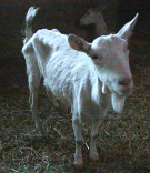
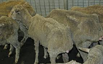
Clinical Signs: This drug resistant bacteria invades the small intestine of an infected animal causing an inflammatory bowel disease resulting in decreased digestion/absorption and severe watery diarrhea, significant weight loss, decreased milk/wool/meat production and premature culling. In sheep and goats, a producer may not see diarrhea but what appears to be signs of parasitism (bottle jaw, anemia, weight/production loss, etc.). When doing fecal exams on animals affected with Johne’s, results indicate parasite resistance to anthelmintic treatment when in fact the animal is immunocompromised which provides a wonderful environment for parasites to take over.
Diagnosis: Serological testing of suspected animals reveals a positive ELISA test. To confirm the positive ELISA test, many veterinarians recommend performing a fecal PCR test which identifies the presence of the bacteria in the animal’s feces allowing contamination of the environment by that animal.
The two primary types of diagnostic tests look for either the organism that causes Johne’s disease (MAP, Mycobacterium avium subspecies. paratuberculosis) or the animal’s response to infection by MAP (antibody in the blood).
Antibody (blood or milk) tests
These assays look for antibody produced by a MAP-infected animal using a technology called ELISA (enzyme-linked immunosorbent assay).

ELISAs are popular because they are fast and the least expensive of the available tests for Johne’s disease. However, they are designed for rapid, low-cost screening of large numbers of animals. ELISAs are less sensitive than MAP-detection assays (PCR and culture), typically being positive in roughly 30%-50% of the animals that MAP-detection assays will identify as MAP-infected. This is generally because antibody production occurs later in the course of a MAP infection, months or even years after an infected animal has been passing MAP bacteria in its feces.
In sheep and goats many present as “poor doers” that may or may not have loose stools. In cattle most positive Johnes disease cattle have extremely loose projectile feces and significant weight loss. The cause for this is that the bacteria invade the epithelial tissue of the intestinal tissue in the large intestine decreasing the ability of the animal to absorb essential nutrients and water.
Treatment
There's no treatment for Johne's disease. However, herd and flock cleanup plans can reduce the prevalence of infection in your animals and eventually eliminate the disease from your herd or flock.
Managing infections can take time. The bacteria that causes Johne's disease can survive for up to a year in the environment, contaminating pastures. Control measures may include:
Tilling the ground or grazing with non-replacement feeder animals.
Test all animals in the herd.
Identify and remove or separate all test-positive animals.
Culling or separating offspring of infected mothers as soon as possible.
Control/Prevention: Control is easy, Don’t Bring in Infected Animals. Try to purchase animals from a Johne’s free herd or at least purchase an animal that tests negative out of a negative dam. After performing the initial test, retest after the quarantine period.
Here are some basic prevention strategies:
Because of the susceptibility of lamb/kids to MAP infection, it is important to keep them well away from manure pellets that may harbor the infection at least for the first 6 months of life, the “window” of maximum susceptibility.
Colostrum, the antibody-rich milk produced by goats in the first few days after giving birth, also can contain MAP. Because colostrum is critical to the health and survival of newborns, feeding colostrum must be done. However, the risk of transmitting MAP infections in colostrum can be minimized by following these three simple rules:
Use colostrum from Johne’s test-negative animals only.
Do not pool colostrum from multiple animals.
Thoroughly wash and dry the udder and teats before collection of aseptic collection of colostrum into clean containers.
Basic Prevention/Control Procedures:
1. Take precautions to avoid introducing infected animals into your herd.
Keep a closed herd or require that replacement animals come from test-negative herds.
Buy animals from low-risk herds.
2. Protect newborns and young animals from coming into contact with contaminated manure.
Have separate birthing areas and keep them clean.
Use different equipment to handle feed and move manure.
Make sure water sources are clean.
When possible, raise young animals in areas separate from adult animals and their manure.
3. Manage colostrum and milk.
Use colostrum from Johne's-negative animals.
Don't pool colostrum.
Clean the udder and teats before collecting the colostrum.
Whenever possible, feed an artificial milk replacer or pasteurized milk instead of raw milk.
Never feed pooled milk or waste milk.
Work with your veterinarian to develop a plan for Johne's prevention and control at your farm.
Resource Links:
Dr. Beth Johnson is a Staff Veterinarian in the Kentucky Department of Agriculture and has 40 years of experience raising and treating small ruminants. Her family farms in Parksville, KY where she raises Gelbvieh cattle and Boer goats.


Comments